Bin Li1 ![]() ,
Hanjun Ma1,
Wenbo Zhang2,
,
Hanjun Ma1,
Wenbo Zhang2,
For correspondence:- Bin Li Email: histlb@126.com
Received: 15 May 2015 Accepted: 11 December 2015 Published: 29 January 2016
Citation: Li B, Ma H, Zhang W, Physicochemical characterization of inclusion complex of catechin and glucosyl-ß-cyclodextrin. Trop J Pharm Res 2016; 15(1):167-172 doi: 10.4314/tjpr.v15i1.23
© 2016 The authors.
This is an Open Access article that uses a funding model which does not charge readers or their institutions for access and distributed under the terms of the Creative Commons Attribution License (http://creativecommons.org/licenses/by/4.0) and the Budapest Open Access Initiative (http://www.budapestopenaccessinitiative.org/read), which permit unrestricted use, distribution, and reproduction in any medium, provided the original work is properly credited..
Purpose:To investigate the suitability of glucosyl-β-cyclodextrin (G-β-CD) to form inclusion complex with catechin, and characterize the physicochemical properties of the inclusion complex of catechin and G-β-CD.
Methods:Catechin and G-β-CD was mixed in water at the same molar ratio, stirred at 20 °C for 48 h and lyophilized to obtain the complex. Its physicochemical properties were investigated by ultraviolet-visible spectrometry (UV), Fourier transform infrared spectroscopy (FT-IR), scanning electron microscopy (SEM), X-ray diffractometry (XRD) and differential scanning calorimetry (DSC).
Results: The characteristic UV absorption peaks for catechin, the physical mixture and the complex occurred at 279 nm. There was no significant difference between the IR spectra of the physical mixture and the complex. SEM and XRD data indicate that catechin was molecularly distributed in G-β-CD matrix and lost its crystallinity in the process. DSC indicate that the heat stability of catechin was significantly improved by complexing with G-β-CD.
Conclusion:Catechin can efficiently interact with G-β-CD to form a complex by freeze-drying method. The complex of catechin and G-β-CD resulted in the changes in some of the characteristic spectral and thermal properties of the former. Furthermore, the heat stability of catechin is significantly improved.
Introduction
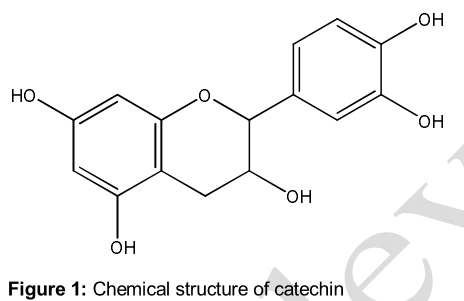
Catechin () is a natural flavonoid and one of the principal active components of green tea, which possesses strong antioxidant, anti-inflammatory, neuroprotective and anticancer activities and so on [1-3]. Therefore, catechin is emerging as a promising bioactive ingredient with potential applications in functional foods and medicines. However, catechin is sparingly soluble in water, which limits its wide applications.
There are some methods that can be used to improve the water solubility of bioactive molecules, such as chemical and enzymatic modification, micro-encapsulation and complexation with cyclodextrins (CDs) [4,5]. Among these methods, complexation with CDs is thought to be a cheap and practical method. β-CD containing seven glucopyranose units is the most common CD [6]. Glucosyl-β-cyclodextrin (G-β-CD) is a glucosyl β-cyclodextrin derivative, with an internal cavity size similar to that of β-CD but is more water-soluble than the native β-CD [7]. However, there are very few studies exploring its suitability for forming inclusion complexes with bioactive compounds.
The objective of this study was to investigate the suitability of G-β-CD as an entrapping agent for forming inclusion complex with catechin, and characterize the physicochemical properties of the inclusion complex of catechin and G-β-CD by ultraviolet-visible spectroscopy (UV), Fourier transform infrared spectroscopy (FT-IR), scanning electron microscopy (SEM), x-ray diffractometry (XRD), and differential scanning calorimetry (DSC).
Methods
Materials and chemicals
Catechin (purity > 95 %) was obtained from Aladdin (Shanghai, China). while G-β-CD (purity 99 %, MW1297) was purchased from Seebio Biotech, Inc. (Shanghai, China). Other chemicals used were of analytical grade.
Preparation of the complex of catechin and G-β-CD
Catechin (0.29 g) and G-β-CD (1.297 g) were dissolved in 25 mL of distilled water, stirred at 20 °C for 48 h and then filtered through a 0.45 μm membrane filter. The filtrate was lyophilized and collected as the complex of catechin and G-β-CD.
Preparation of the physical mixture of paeonol and G-β-CD
Catechin (0.29 g) and G-β-CD (1.297 g) were mixed and pulverised in ceramic mortars. The obtained powder was used as the physical mixture of catechin and G-β-CD.
UV spectroscopy
UV absorption spectra were recorded for catechin, G-β-CD, their physical mixture and the inclusion complex by using a UV recording spectrophotometer (Purkinje, Beijing, China). Each sample was dissolved in water at the room temperature. The aqueous solutions were scanned, respectively, in the range 220 to 400 nm to obtain the UV absorption spectra.
Fourier transform infrared spectroscopy (FT-IR)
The FT-IR spectra of catechin, G-β-CD, their physical mixture and the inclusion complex were collected between 4000 and 400 cm-1 (Mid infrared region) on a TENSOR 27 FT-IR spectrophotometer (Bruker, Germany) with 256 scans at a resolution of 4 cm-1. Each sample was ground with spectroscopic grade potassium bromide (KBr) powder and then pressed into 1 mm pellets (2 mg of sample per 200 mg dry KBr). A blank KBr disk was used as background. FT-IR spectra were the OPUS software (Bruker, Germany).
Scanning electron microscopy (SEM)
The surface morphology of catechin, G-β-CD, their physical mixture and the inclusion complex was examined by a Quanta 200 environmental scanning electron microscope (FEI, USA). Prior to examination, samples were prepared by mounting about 0.5 mg of powder onto a 5 mm × 5 mm silicon wafer affixed via graphite tape to an aluminium stub. The micrographs were obtained with an accelerating potential of 20 kV under low vacuum.
X-ray diffractometry (XRD)
According to a previous method [8], the X-ray powder diffraction patterns of catechin, G-β-CD, their physical mixture and the inclusion complex were obtained with a D8 Advance X-ray diffractometer (Bruker, Germany) using a Ni-filtered, Cu Ka radiation. All samples were measured in the 2θ angle range between 5 and 80o.
Differential scanning calorimetry (DSC)
According to a previous method [9], DSC analysis was carried out for catechin, G-β-CD, their physical mixture and the inclusion complex with a Q200 differential calorimeter calibrated with indium (TA Instruments, USA). Each powder (3 mg) was heated in a crimped aluminium pan at a scanning rate of 10 °C/min between 30 and 280 °C temperature range under a nitrogen flow of 40 mL/ min. An empty pan sealed in the same way was used as reference.
Results
UV spectra
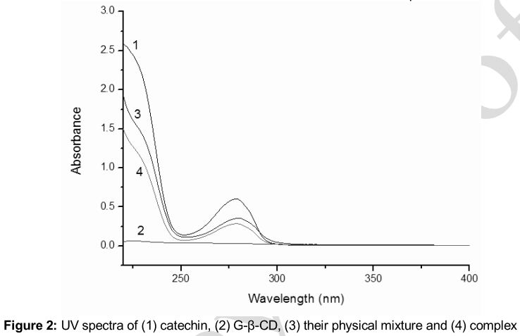
The UV spectra of catechin, G-β-CD, their physical mixture and the inclusion complex were shown in . There was no UV absorbance of G-β-CD in the range of 220 to 400 nm. There was also no difference between the UV spectra of the physical mixture and the complex. The characteristic absorption peaks of catechin, the physical mixture and the complex were still present at 279 nm.
IR spectra
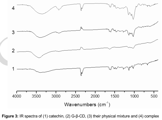
The IR spectra of catechin, G-β-CD, their physical mixture and the inclusion complex were shown in . The IR spectra of the complex and the mixture of catechin and G-β-CD were similar to that of G-β-CD for the low content of catechin in the system. And there was no significant difference between the physical mixture and the complex.
Particle morphology
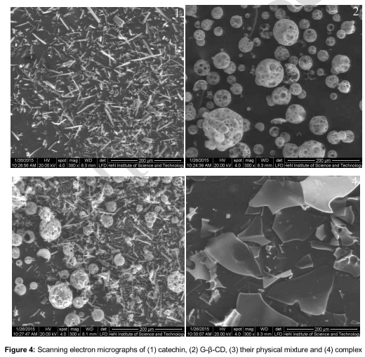
The scanning electron micrographs of catechin, G-β-CD, their physical mixture and the inclusion complex were shown in . Catechin existed in needle-like crystal while G-β-CD was observed as amorphous spheres. For the physical mixture, both the needle-like crystals and amorphous spheres could be observed. In contrast, the catechin/G-β-CD inclusion complex appeared in the form of irregular particles.
XRD diffractograms
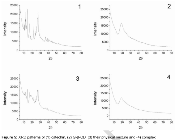
The XRD patterns of catechin, G-β-CD, their physical mixture and the inclusion complex were shown in . The XRD pattern of catechin displayed sharp crystalline peaks while the XRD pattern of G-β-CD revealed a broad peaks in the range of range of 15-25o. Compared with the XRD pattern of the physical mixture, the crystalline peaks had disappeared in the XRD pattern of the complex.
Thermal characteristics
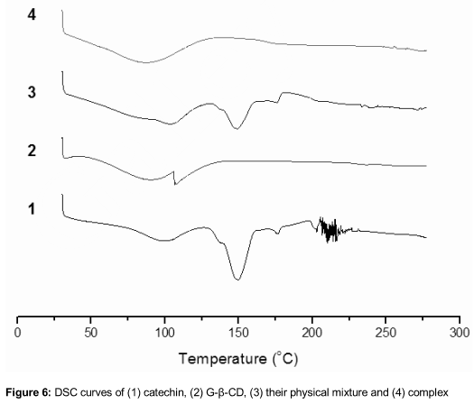
The TG curves of catechin, G-β-CD, their physical mixture and the inclusion complex were shown in . Catechin displayed one big endothermic peak, corresponding to the melting point of the crystalline form. And a broad endothermic peak of G-β-CD was observed at about 100 °C for the loss of water. The DSC curve of the physical mixture mainly showed the individual characteristics of catechin and G-β-CD. For the complex, the disappearance of the melting peak of catechin could be found.
Discussion
β-CD is most widely used to improve the aqueous solubility and stability of bioactive molecules. However, the aqueous solubility of β-CD in water is only 1.85 g/100 ml at 25 °C) [10]. As a result, some β-CD derivatives have been developed. G-β-CD is prepared by enzymic method, whose solubility in water was significantly higher than that of β-CD [11].
In this study, the freeze-drying method was used to prepare the inclusion complex of catechin and G-β-CD. UV and IR results showed that catechin and G-β-CD in the complex interacted by non-covalent bond. SEM demonstrated that when the solutions of the two compounds were freeze-dried, they formed a close association, probably in the form of inclusion complex, in which catechin and G-β-CD no longer exist in their native states. XRD also proved that catechin in the complex was molecularly dispersed in G-β-CD matrix.
For DSC, The physical mixture exhibited the combined characteristics of both molecules, indicating that no close association formed between the two molecules when the two powders are simply mixed together. In contrast, the DSC curve of the inclusion complex exhibited mainly the features of the G-β-CD curve while the characteristic endothermic peaks of catechin disappeared entirely, which suggests that an association structure was formed between the two molecules and that the heat stability of catechin was significantly improved [12].
Conclusion
The results of this study demonstrate that catechin can be efficiently complexed with G-β-CD to form an inclusion complex by freeze-drying method. The complex formed has different physicochemical characteristics from that of the free catechin. Thus, complexation with G-β-CD is a promising strategy to improve the application of catechin in functional foods and medicines.
Declarations
Acknowledgement
References
Archives


News Updates